Scanning Electron Micrographs Trap Jaw Ant
Feb 2, 2013 18:31:02 #
eframgoldberg
Loc: South Florida
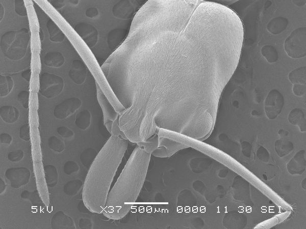
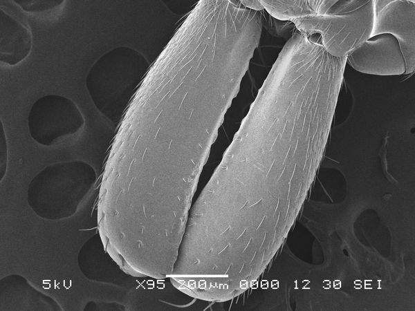
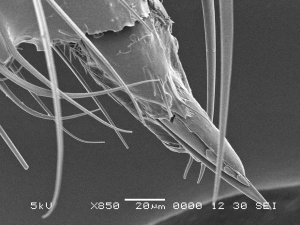
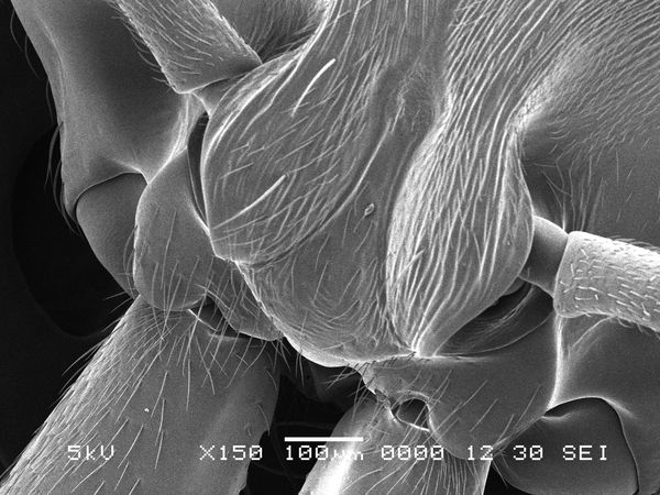
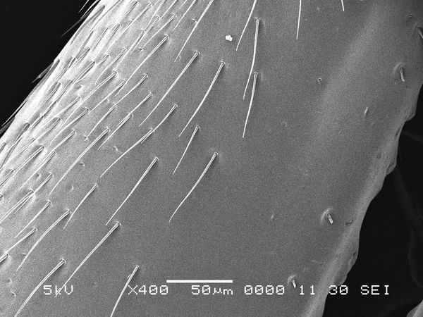
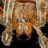
SEM images of a Trap Jaw Ant Odontomachus

Stinger
mandibles attached to head
mandibles 400x
Feb 2, 2013 19:17:01 #
eframgoldberg wrote:
SEM images of a Trap Jaw Ant Odontomachus
Pretty cool! Thanks for sharing.
Feb 2, 2013 20:39:55 #
Feb 3, 2013 05:40:38 #
eframgoldberg
Loc: South Florida
Yes, I have access to the one in the biology department here at FIT
Feb 3, 2013 05:41:02 #
Feb 3, 2013 12:07:46 #
While attending BIP in the late 70s, I had (clandestine) access to an ESM at UCSB. We scanned individual diatoms, separated from diatomaceous earth. Our imprinted scale was in microns.
Feb 3, 2013 13:27:11 #
eframgoldberg
Loc: South Florida
yeah the scale on these photos are in micrometers, diatoms and pollen are great since you need such high magnification to see them in the first place
Feb 3, 2013 14:17:49 #
eframgoldberg wrote:
Yes, I have access to the one in the biology department here at FIT
Just curious. What are you studying?
Feb 3, 2013 14:27:39 #
eframgoldberg
Loc: South Florida
Getting my PhD Chemistry, but I went to the SEM technician and showed her my photographer/photo stacking and she has given me access to the microscopy department
Feb 3, 2013 15:35:39 #
eframgoldberg wrote:
Do you focus stack these microscopic images? Can't imagine the challenge in that!I went to the SEM technician and showed her my photographer/photo stacking and she has given me access to the microscopy department
Feb 3, 2013 23:47:08 #
eframgoldberg
Loc: South Florida
nope, the magic behind scanning electron microscopy is large depth of field at high magnifications :)
Feb 4, 2013 02:39:37 #

Really nice images and thanks for sharing... But!, I agree with Douglass... Not Fair!...
:( :(
:( :(
Feb 4, 2013 10:33:15 #
eframgoldberg
Loc: South Florida
LoneRangeFinder wrote:
From what I understand and I might be wrong, a photograph shot with a lens and light involves focusing the rays onto a plane. The rays converge into spots or airy disks and between a certain diameter they are considered in focus or out of focus. Due to physics there are limitations on the relationship between resolution, magnification, and depth of field. Do you focus stack these microscopic images? Can't imagine the challenge in that!
A scanning electron microscope can work several ways but basically a stream of electrons is shot at a sample and this bounces electrons off the sample which are then detected. In this way it functions more like a 3 dimensional scanner, giving a large depth of field.
Also while visible light is confined to about 400nm to 700nm, the electrons in an SEM are accelerated very quickly giving wavelengths up to 1000 times smaller. This allows for even higher magnification than is possible with visible light.
Feb 4, 2013 13:19:29 #
An SEM works much like an X-ray machine, in that much is in focus on final image. Deep DoF.
Feb 5, 2013 10:11:01 #

If you want to reply, then register here. Registration is free and your account is created instantly, so you can post right away.