Posts for: James Shaw
Mar 4, 2017 16:50:56 #
CathyAnn wrote:
Excellent shot! 
Thanks, CathyAnn. Appreciate the comment!
Mar 4, 2017 13:48:40 #
Swamp-Cork wrote:
James, I'm attempting to sent the article but not so great on sending such things on the computer and if it doesn't make it through just send a message and I will snail mail it to you Corky
Got it, and with some manipulations with another program, I was able to read it clearly. Thank you. Nice article with many facts about insects that are new to me. Just a little scary about the declining populations of so many insects, but the author left hope of their return with time and help from back yard planters, like me, and by others who plant along the roadways.
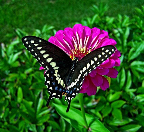
I have planted milkweed in my yards for Monarchs, and help other butterflies, which pass through during the changing seasons and visit my annually-planted bed of zinnias (see some visitors that I captured below). I love capturing them with my camera when they visit
Thank you, again, for the great article and for your nice comments and other information that you provided. It has been such a nice education for me.
- Jim

Mar 4, 2017 10:48:57 #
Swamp-Cork wrote:
James, There was an excellent article published i... (show quote)
Yes, I would like to read the article, but looked on-line, and it requires that one subscribe to the magazine, of which I am not a subscriber. Your Virginia is a most beautiful state with abundant and various forms of wildlife. If I were to receive the article, I would enjoy reading it for sure.
Mar 3, 2017 19:51:38 #
relbugman wrote:
Fascinating, indeed! Thanks, again, for another interesting facet regarding another of Mother Nature's beauties and its activities. Can't wait to see them, once again, in action this Spring, on my wood deck and fence. I have noticed they will sometimes bore right into my pressure-treated-deck wood, but don't recall seeing anything emerge. Maybe they were poisoned by the heavy metals.Just a quick correction: Carpenter bee adults do ... (show quote)
Mar 3, 2017 11:41:28 #
Swamp-Cork wrote:
Same here. Honey bees have almost disappeared. I used to be careful walking barefoot on clover but in the past several years almost no honey bees at all. Hopeful they will return to pollinate and carry on as they used to do in such abundance. For some reasons, unknown to me, their numbers have been greatly reduced. Perhaps those now selected and still living will repopulate the honey bee population and spread their resistant genes.Actually, I believe that I see more carpenter bees... (show quote)
Mar 2, 2017 09:16:01 #
relbugman wrote:
Well, you’ve collared both me and my college ex-ro... (show quote)
A most scholarly and really interesting feedback, relbugman. With an undergraduate Biology degree, from so many years ago, I was able to follow the details of your most interesting post, and I read it all! Thank you for your thoughtful and carefully detailed response. Especially enlightening is that there was no final exam at the end. I never liked final exams and had sympathy for those having to take the finals I had to give them. Final exams often enter my dreams and haunt me, as in the dreams I have not attended class in some time and am on the way to the final, unprepared. I wake up sweating.
Again, thanks so much for contributing to my continued education!! Matter now settled, and I thank all others who participated in this post, as well. Most informative. Education never ends or should never end.
Mar 1, 2017 20:06:46 #
docshark wrote:
Thanks so much for your input, docshark. I shall now look for it on dragonflies. And much appreciate your positive comments on my photo. I have learned a lot here.Well James, you will find that membranous attachment on many insects. Many of my dragonfly photos show it quite nicely as a dragonfly must be able to move it's head in all directions for protection and to hunt. Unfortunately the structure has no real name. As you stated it does encase the nerves controlling the mouthparts and eyes. Excellent shot by the way. Nice color and composition. Have a great day!
-Doc
-Doc
Mar 1, 2017 11:59:33 #
merrytexan wrote:
beautiful shot james!
Merrytexan, much thanks for your kind words!
Mar 1, 2017 11:57:22 #
sailorsmom wrote:
What a beautiful shot, James!
Thank you sailorsmom for you nice words!
Mar 1, 2017 11:55:30 #
Swamp-Cork wrote:
Great image and we have many in our area!
Yes, and Virginia is not that far away. Probably will see many more this Spring. Beautiful creatures. And thanks for the comment.
Mar 1, 2017 11:54:01 #
James Shaw wrote:
Any etymologists on UHH? If so, do you know what the ligament-like strands are called, shown between the head and thorax, that allow the bee's head to bend away from its thorax?
CORRECTION:
It was rightfully brought to my attention by an UHH member (who kindly communicated by private message) that the use of the word "etymologists" in my original post (above) was incorrect. I should have used the word, "entomologists." My apologies for the mistake.
I am of the school: "Live and Learn and Pass It On"
Mar 1, 2017 11:34:38 #
Mr. B wrote:
Here's a diagram of insect morphology. Copyright of Piotr Jaworski and used with permission. As to the "joint" I would think it's some kind of chitinous joint. It is, after all, an exoskeleton. Maybe some one else can provide enlightenment.
Simply gorgeous insect diagram, on download. Thank you! Your offer of "chitinous joint" is the best yet that I have gotten for an explanation for the structure that I see. One would think that there are bundles of nerves encased in that joint that communicate brain and spinal cord. Number 5 on the chart indicates cerebral ganglia, so perhaps that is as close as I can get.
Thank you, again, Mr. B.!
Feb 28, 2017 20:17:20 #
Feb 28, 2017 16:29:39 #
Dixiegirl wrote:
Doc would probably know the answer better than anyone I know, James, and I hope he sees your post. This is such a beautiful image.
Thank you, Dixiegirl.
Feb 28, 2017 16:28:35 #
Nikonian72 wrote:
I photographed this image several Springs back in South Central Ohio. Just got around to post-processing some old photos. I guess I need a bee anatomy book to see the detailed anatomy of bee, neck-to-thorax attachment organs? I shall, now, rename this photo to Xylocopa virginica (Eastern Carpenter bee) - thanks for the name! Just curious, what designates the sex, a "female?"This appears to be a female Eastern Carpenter bee (Xylocopa virginica). Where was it photographed?