Photomicrography
Jul 13, 2013 07:57:37 #
Some of you do photography of space with telescopes, and many do macros. Do any of you take pictures through microscopes?
Jul 13, 2013 08:58:01 #
I have been doing photomicrography ever since high school in 1969 when I built a system using a highly modified Kodak Folding Ensign camera that belonged to my grandfather and a not quite so venerable Olympus GB. It was for a science fair project. I learned how to develop film and make prints for that. Evolved through a Zeiss photomicroscope at university with Panatomic X and a 16 mm Bolex movie camera with Tri- X reversal to VCR and now a Pixera 5.8 Mpx cooled chip camera and Olymous BX50 microscope and streaming video capture. I now teach a postgrad course in light microscopy and digital image processing. With retirement a few years away I have my my eye on a D600 with 105 mm macro lens and R1 flash kit. There's a wonderful world down there...
Jul 13, 2013 09:03:05 #

jerryc41 wrote:
Some of you do photography of space with telescopes, and many do macros. Do any of you take pictures through microscopes?
There is a very seldom contributor in the UHH Macro section with access to a research electron microscope at a Medical university. He has submitted some amazing images--but I cannot recall his name. I'll see if I can locate his last post.
Jul 13, 2013 12:28:44 #

Jul 13, 2013 12:50:22 #
CSI Dave
Loc: Arizona
LoneRangeFinder wrote:
There is a very seldom contributor in the UHH Macro section with access to a research electron microscope at a Medical university. He has submitted some amazing images--but I cannot recall his name. I'll see if I can locate his last post.
It's not me, but I often use our SEM. However, my purpose is usually not to get a pretty picture, but to get elemental data from the electron microscope.
I use optical microscopes with a digital camera all the time. I should go through the pics and see if there's anything worth posting.
Jul 13, 2013 13:07:59 #

Jul 13, 2013 22:04:17 #

I do, especially photos of diatoms using Nikon D300s on vintage Zeiss photomicroscope and Nikon LKE microscopes. I am still tying to perfect my techniques.
Jul 13, 2013 22:18:32 #
silveragemarvel wrote:
I do, especially photos of diatoms using Nikon D300s on vintage Zeiss photomicroscope and Nikon LKE microscopes. I am still tying to perfect my techniques.
I always thought of the, as looking like little snowflakes.
Jul 13, 2013 22:29:29 #
Diatoms are the best. Beautiful,easy to collect, full of details that increase with increasing magnification, and they are well documented. They were once used as reference specimens for testing resolution. Rheinberg illumination can show them off in spectacular fashion.
Jul 13, 2013 22:32:51 #
bioteacher
Loc: Brooklyn, NY
LoneRangeFinder wrote:
There is a very seldom contributor in the UHH Macro section with access to a research electron microscope at a Medical university. He has submitted some amazing images--but I cannot recall his name. I'll see if I can locate his last post.
The specimen has to be dead in order to use an electron microscope.
Jul 14, 2013 06:10:39 #

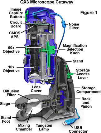
I have an Intel-Mattel Microscope, which was sold as a toy by many in the 1990s. While compared to today's camera resolution it was low, it was quite sufficient for me to have a client in Australia to send metal etched surfaces photos to me, USA, for comments. The unit sold for less than $100 and were world wide so they were able to purchase one in the land down under.
http://micro.magnet.fsu.edu/optics/intelplay/intelanatomy.html
There is a thread that your question lead me to find, thank you. In this thread it discusses the use on InfarView with TWAIN interface to view and control the microscope rather than the QX3 software. http://www.lencoheaven.net/forum/index.php?topic=620.0
There are other microscopes (low cost) out there. A truly different approach from the use of reversed lenses as macro image producers.
In the 70's I did a fair amount of work evaluating a number of candidate SEMs for Firestone Fibers (part of Tire company). I was so good at evaluating them that management felt that the evaluation photos solved the problem and we did not get a SEM. I was disappointed,, what a fascinating Toy for a Kid like me!!
Thank you again for lighting a fire under my Intel/Mattel Scope and Me. I just bought one on E-Bay for less than $20 total. I plan to take it apart and perhaps replace the sensor portion with that from a low cost multi megpix camera... gotta have some fun!
MATTEL-INTEL MICROSCOPE DISECTED
Jul 14, 2013 06:57:58 #
dpullum wrote:
I have an Intel-Mattel Microscope, which was sold ... (show quote)
Thanks for that interesting story.
Jul 14, 2013 07:21:26 #
Pablo8
Loc: Nottingham UK.
I still have my Nikon microscope, with film camera body, built-in-flash, and bino' stereo viewers. I used to do crystal formations with polarized illumination/and or dark-field illumination. Wonderfull 'Toy' ...Must try it out again with digital camera body attached this time.
Jul 14, 2013 07:26:08 #
I'm involved in a book project requiring studio shots using telephoto lenses down to ultramicro. Many of the macro shots have a field of view of less than 1 cm wide, but sometimes the entire view on the Canon 60D sensor has 1mm width of field. I used to photograph through regular microscopes, but with digital photography I'm able to convert my camera into a microscope. Since the 1950's, the lens threads of microscope lenses have been standardized to Royal Microscopical Society pitch and diameters [RMS] (20.5 mm). Using microscope objectives is a growing segment of macrophotography. I just got a Cambridge Instruments 50X ULWD lens to try out. It turns out it will be a trial by fire as I'm taking it to a photo shoot this week. ULWD means ultra long working distance. In this case, the microscope lens has an astonishing 2 cm working distance. It also has a 2 micron depth of field. That means that stacking images of a thin blond hair would require 20 images minimum to show an in-focus picture of the side view. For larger objects, 100-500 microns, I use a 25mm Luminar microscope objective, which also has a 2 cm working distance from glass to object. The obvious problem is that when you are using reflected light, rather than transmitted, the lens can get in the way. One microscope lens I tried had so little working distance that the lens was virtually touching the object (well some large number of microns from it). The side light was still diffracted as the separation acted as a slit and the illuminated surface from a white-light balanced source resulted in a dark foggy red brown haze as the best illumination possible. At any rate, using suitable microscope objectives directly on a camera bellows or extension rings offers better opportunity for versatility and sharpness than shooting through a conventional microscope. Working distance is the limiting factor. A friend of mine in Japan posted wonderful close-ups he got with a 16mm Luminar lens, but then sadly reported he had damaged two very important specimens because his lens had very little working distance and it crunched into then. The glass survived unscratched.
Jul 14, 2013 07:39:32 #
newryqs wrote:
I'm involved in a book project requiring studio sh... (show quote)
It's a whole other world!
If you want to reply, then register here. Registration is free and your account is created instantly, so you can post right away.